
A Celebration of the Beauty of Cells
Beautiful Cells
Ok, not everyone would look at an image of a cell and see something beautiful, but I guess that explains why I love biology. In a previous blog titled “Innovative Products Featured at the ASCB Conference Part I,”I discussed some of the interesting products and innovations that I saw at the Annual Meeting of the American Society for Cell Biology (ASCB) in San Francisco. As mentioned in the blog, one display that was impossible to ignore was GE Healthcare’s beautiful cell images presented in high definition color as part of their Annual Cell Imaging Competition. The competition, in its sixth year, received over 100 images entered for the high-content analysis and high and super resolution microscopy categories. Images were submitted from researchers who are “investigating at the cellular level conditions such as cancer, HIV, and neurodegenerative disease”. After an Expert Scientific Panel selected the top 15 images in each category, the public was able to vote on their favorites both at the conference and online.
GE Healthcare Life Sciences Cell Imaging Competition
Yesterday, GE Healthcare Life Sciences announced the three overall winners of the 2012 Cell Imaging Competition. The winners will have their images displayed in Times Square on NBC Universal’s HD Screen at a special event on April 19-21, 2013. GE Healthcare generously allowed us to also post the winning images on The Cell Culture Dish. I hope you enjoy the winning images as much as I did. I am so pleased that the images will also be displayed in Times Square, hopefully inspiring budding scientists to pursue a career in life sciences.
In a press release issued by GE Healthcare, Eric Roman, General Manager of Research and Applied Markets, GE Healthcare Life Sciences, said: “This year’s winning images are as beautiful and compelling as ever. Not only can they be appreciated from an aesthetic point of view, but they remind us of the cellular complexity behind disease and why the study of cells is so important. We were delighted to receive so many outstanding entries to the competition, which highlights how high-content and super-resolution cell imaging are helping scientists explore the universe of the cell, and so advance our understanding of so many life threatening and life-limiting diseases.”
The winning images and gallery of the 2012 Cell Imaging Competition are available at www.gelifesciences.com/cellimagecompetition. If you are interested in submitting your beautiful cell images, the competition will be held again in 2013.
Winning Images:
1st place – High and Super Microscopy category
Jane Stout, School of Medicine, Indiana University, Bloomington, USA
Image Description: Metaphase epithelial cell stained for microtubules (red), kinetochores (green) and DNA (blue).
Disease focus: Cancer

Winner: Microscopy
Photo by: Jane Stout
1st place – High-Content Analysis category
Anushree Balachandran, Genea, Sydney, Australia
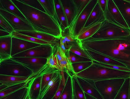
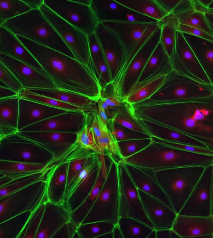
Image description: Huntington’s stem cell derived oligodendrocyte precursors stained for phalloidin (green), vinculin (red) and DNA (blue).
Disease focus: Huntingdon’s disease
Winner: High-Content Analysis
Photo by: Anushree Balachandran
Regional Winner – Microscopy category
Markus Posch, Centre for Gene Regulation and Expression, University of Dundee, UK
Image description: Prometaphase human cervical carcinoma (HeLa) cell with GFP-histone labeled chromosomes (blue) stained for tubulin (yellow).
Disease focus: Cancer

Winner: Regional
Photo by: Markus Posch