
Captivating Cell Images
Cell Images
“Cells are beautiful,” it is a statement that I made last year when I posted a blog on the winners of GE Healthcare’s Annual Cell Imaging Competition and once again I am reminded how true it is. Cell images are certainly captivating and fun to look at, but what these images can tell us about cell health and function, disease states, and the impact of possible treatments can be ground-breaking. The competition, in its seventh year, received over 100 images entered for the high-content analysis and high and super resolution microscopy categories. Images were submitted from researchers who are using cell imaging to look at a wide variety of therapeutic areas.
GE Healthcare Life Sciences Cell Imaging Competition
Last week, GE Healthcare Life Sciences announced the three overall winners of the 2013 Cell Imaging Competition. The winners will have their images displayed in Times Square at a special event between April 25-27, 2014. GE Healthcare allowed us to also post the winning images on The Cell Culture Dish. I hope you enjoy the winning images as much as I did. I am so pleased that the images will also be displayed in Times Square, hopefully inspiring budding scientists to pursue a career in life sciences.
In a press release issued by GE Healthcare, Eric Roman, General Manager of Research and Applied Markets, GE Healthcare Life Sciences, said: “ This year’s three winning images are once again incredibly beautiful and compelling, reminding us of the cellular complexity behind disease and why the study of cells is so important.”
The winning images and gallery of the 2013 Cell Imaging Competition are available at www.gelifesciences.com/cellimagecompetition. If you are interested in submitting your beautiful cell images, the competition will be held again in 2014.
Winning Images:
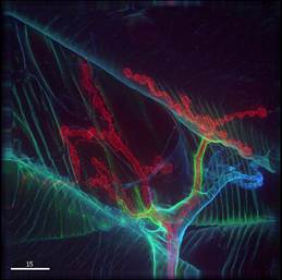
1st place – Microscopy category
Vanessa Auld, University of British Columbia, Vancouver, Canada.
Image Description: Drosophila neuromuscular junction stained for extracellular matrix proteins (green and blue) and the nerve terminal (red).
Therapeutic focus: Neurodegenerative disease.

Winner: Microscopy
Photo by: Vanessa Auld
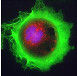
1st place – High-Content Analysis category
Martin Barr, St James’s Hospital and Trinity College Dublin, Dublin, Ireland.
Image description: Lung adenocarcinoma cell stained for F-actin (green), mitochondria (red) and DNA (blue).
Therapeutic focus: Cancer.
Winner: High-Content Analysis
Photo by: Martin Barr
Regional Winner – Microscopy category
Graham Wright, Institute of Medical Biology, A*STAR, Singapore
Image description: Mouse spermatocyte spread stained for KASH-5 and SCP3 (red and green) and DNA (blue).
Therapeutic focus: Fertility

Winner: Regional
Photo by: Graham Wright