Cell Culture and Single Cell Passaging of Human Pluripotent Stem Cells Without the Need for ROCK Inhibitor
Corning® PureCoatTM rLaminin-521 Cultureware: A Ready-to-use, Animal-free Vessel Platform for the Culture and Single Cell Passaging of Human Pluripotent Stem Cells
A guest blog by Himabindu Nandivada, Jorge A. Montoya, and Deepa Saxena Corning Incorporated, Life Sciences Bedford, Massachusetts
Introduction
Human pluripotent stem cells (hPSCs), including human embryonic stem cells (hESCs), and human induced pluripotent stem cells (hiPSCs) have the ability to self-renew and to give rise to specialized cell types and therefore, have tremendous potential in clinical, drug discovery, and regenerative medicine applications¹. Human PSCs are traditionally cultured on a layer of feeder cells (such as mouse or human fibroblasts)². For feeder-free cultures, these cells are cultured on either complex mixtures of naturally- derived extracellular matrices (such as Corning Matrigel® matrix), or more recently on a variety of recombinant proteins (such as Laminin) and synthetic substrates (such as Corning Synthemax® II-SC)².
Human PSCs are typically propagated as colonies and passaged by clump passaging techniques (i.e., cutting colonies into small clumps or clusters for passaging) which is labor intensive and adds to variability in cell seeding³. Alternatively, culture of dissociated hPSCs as a single cell suspension requires the utilization of small molecules such as Rho-associated protein kinase (ROCK) inhibitors (e.g., Y-27632 or thiazovivin) to prevent dissociation-induced apoptosis4. The use of completely dissociated single cell suspension for passaging generates a monolayer culture that has advantages of higher culture scalability, rapid expansion, and high efficiency5. However, culture conditions which involve single cell dissociation of hPSCs have been shown to cause genomic alterations in hPSCs during long-term cultures6,7.
Recently, a recombinant isoform of human Laminin, namely Laminin-521, has been shown to support long-term single cell culture without the need for ROCK inhibitors8-12. This work has been performed using surfaces freshly-coated with rLaminin-521. However, manual coating procedures are labor-intensive and can introduce variability into the culture environment.
Here we present Corning PureCoat rLaminin-521 cultureware, a sterile pre-coated, ready-to-use, animal-free surface manufactured under cGMP conditions that enables stem cell scientists to efficiently scale-up their cell production in xeno-free culture environments.
PureCoat rLaminin-521 cultureware supported long-term culture, and single cell passaging of hiPSCs in xeno-free NutriStemTM XF/FF culture medium. The hiPSCs remained undifferentiated as demonstrated by the expression of OCT-3/4 and SSEA-4 markers. Cells maintained pluripotency through 10 passages and successfully differentiated into cells from the three germ lineages. Human iPSCs possessed a normal karyotype after 10 passages on PureCoat rLaminin-521 cultureware as a single cell suspension without the use of ROCK inhibitor. PureCoat rLaminin-521 cultureware removes manufacturing bottlenecks associated with self-coating protocols with respect to time, labor, and coating variability, and provides a robust and scalable platform for hPSC culture.
Materials and Methods
Human induced pluripotent stem cell (hiPSC) culture and passaging on Corning Matrigel matrix
Human episomal iPSC line (Thermo Fisher Scientific) was used for this study and was maintained in feeder-free conditions on Corning Matrigel hESC-qualified matrix (Corning Cat. No. 354277) in mTeSR™1 medium (STEMCELL Technologies) according to manufacturers’ instructions. To passage cells on Matrigel matrix, culture medium was aspirated and cells were washed once with DPBS (2 mL/well; without calcium and magnesium). Cell dissociation buffer (1 mL/well; Enzyme-free, PBS; Thermo Fisher Scientific) was added to the wells and incubated for 3 to 5 minutes at room temperature. The separation of the cells within the colonies was observed under the microscope. The cell dissociation buffer was carefully aspirated from the wells, and the cells were dislodged from the plate as small cell clumps/clusters by forcefully adding fresh medium to the well. The small cell clumps/clusters were seeded onto a freshly-coated Matrigel matrix plate at a ratio of 1:10 to 1:14 in mTeSR1 medium (2 mL/well) and incubated at 37°C and 5% CO2 in a humidified incubator. The medium was changed daily and the cells were passaged once a week. The hiPSCs were kept in continuous culture under these conditions until needed for the experiments.
Long-term single cell hiPSC culture on Corning® PureCoatTM rLaminin-521 cultureware
NutriStem XF/FF culture medium (1 mL/well, Stemgent) was added to each well of PureCoat rLaminin-521 cultureware (6-well plate, Corning Cat. No. 356290). These plates were equilibrated in a humidified incubator at 37°C for 30 minutes to 1 hour until the cells were ready to be seeded.
Human iPSCs growing on Corning Matrigel® matrix (or rLaminin-521) coated surfaces were washed once with DPBS (2 mL/well). StemPro® Accutase® cell dissociation reagent (1 mL/well, Thermo Fisher Scientific) was added to the wells and the cells were incubated at 37°C and 5% CO2 in a humidified incubator for 3 to 4 minutes. The plate was gently tapped on the side to further detach the cells from the surface. The cells were gently pipetted up and down 6 to 10 times to achieve a single-cell suspension. Fresh medium (3 to 4 mL/well) was added to the cell suspension to dilute the Accutase, and the cell suspension was centrifuged at 100 × g for 4 minutes. The supernatant was removed and the cells were resuspended in culture medium. The cells were counted using a Vi-CELL™ Cell Viability Analyzer (Beckman Coulter). The hiPSCs were seeded at a density of 50,000 cells/cm² on to the pre-equilibrated rLaminin-521 plates. A complete medium change was performed 48 hours after cell seeding and cells were fed daily until they were ready to be passaged. Cell morphology was observed daily using an EVOS® XL Cell Imaging System (Thermo Fisher Scientific). Cells were passaged every 4 to 5 days when the cell confluence was more than 80%.
Undifferentiated marker expression using flow cytometry analysis
Cells were dissociated from rLaminin-521 plates with Accutase as described above and samples were prepared for FACS according to the manufacturer’s instructions for the BD Stemflow™ Human and Mouse Pluripotent Stem Cell Analysis Kit (BD Biosciences). The cells were analyzed with a BD FACSCalibur™ flow cytometer (BD Biosciences) and data were analyzed with the BD CellQuest™ Pro software (BD Biosciences).
Tri-lineage differentiation
To assess whether the hiPSCs maintained pluripotency, Human Pluripotent Stem Cell Functional Identification Kit (R&D Systems) was used according to the protocols provided. The cells were plated onto Corning Matrigel matrix-coated 24-well plates and differentiated into the three lineages separately. The three germ layers were stained with antibodies for Otx2 (ectoderm marker), brachyury (mesoderm marker), and SOX17 (endoderm marker). The cells were counterstained with the corresponding secondary antibody (Anti-goat IgG-NL557, R&D Systems) and nuclei were labeled with Hoechst 33342 (Thermo Fisher Scientific). The stained differentiated cells were imaged using a fluorescence microscope (Olympus IX70).
Karyotype analysis
The hiPSCs were dissociated with Accutase as described above and seeded on Corning Matrigel matrix-coated T-25 flasks with ROCK inhibitor Y27632 (5 μM, Sigma). Live cell samples were submitted for karyotyping by G-banding analysis (Cell Line Genetics).
Results and Discussion
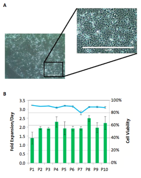
Human iPSCs were cultured on Corning PureCoat rLaminin-521 cultureware in NutriStem XF/FF culture medium for 10 passages. Throughout this study, the hiPSCs exhibited the typical undifferentiated hPSC morphology as characterized by the presence of small cells and high nucleus to cytoplasm ratio (Figure 1A). The cells maintained a high viability (average of 88.9% ±3.4) and cell expansion rates were consistent between passages as evidenced by the fold expansion rate at each passage (Figure 1B).

After 10 passages, hiPSCs remained undifferentiated as demonstrated by the flow cytometry data (Figure 2). Human iPSCs expressed undifferentiated hPSC markers OCT3/4 (>94%) and SSEA-4 (>95%) while the differentiation marker SSEA-1 (0.2%) was not detected.

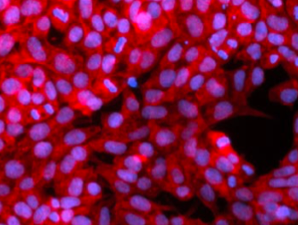
To assess whether the hiPSCs maintained pluripotency in vitro after 10 passages on rLaminin-521 cultureware, the cells were subjected to directed differentiation to the three germ layers, endoderm, mesoderm, and ectoderm, using media supplements that drive differentiation. The differentiated cells were characterized using germ layer markers specific for ectoderm (Otx2), mesoderm (brachyury), and endoderm (SOX17) (Figure 3). The hiPSCs cultured on Corning® PureCoatTM rLaminin-521 cultureware were able to differentiate into the three germ layers demonstrating that rLaminin-521 cultureware supported the expansion of hPSCs without affecting their pluripotency.

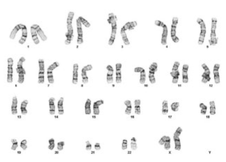
After 10 passages, the cells exhibited a normal karyotype (Figure 4) and genetic abnormalities were not detected.

Conclusions
Corning PureCoat rLaminin-521 cultureware supported the long-term culture and single cell passaging of human pluripotent stem cells in xeno-free NutriStem XF/FF culture medium. The human pluripotent stem cells remained undifferentiated, pluripotent, and possessed a normal karyotype after 10 passages using single cell passaging method without the use of ROCK inhibitor.
Corning PureCoat rLaminin-521 cultureware is a ready-to-use, animal-free, cGMP manufactured, sterile (10-3), nonpyrogenic and functional pre-coated platform available in multiple vessel formats to enable users to scale up their hiPSC culture.
For more information about Corning® PureCoatTM rLaminin-521 cultureware for culture of human pluripotent stem cells , please visit – http://www.corning.com/worldwide/en/products/life-sciences/products/surfaces/rlaminin-cultureware.html
References
- Carpenter MK and Rao MS, Concise review: making and using clinically compliant pluripotent stem cell lines, Stem Cells Translational Medicine (2015), 4(4):381-388.
- Villa-Diaz LG, et al., Concise Review: The evolution of human pluripotent stem cell culture: from feeder cells to synthetic coatings, Stem Cells, (2013), 31(1):1-7.
- Chen KG, et al., Human pluripotent stem cell culture: considerations for maintenance, expansion, and therapeutics, Cell Stem Cell (2014), 14(1):13-26.
- Watanabe K, et al., A ROCK inhibitor permits survival of dissociated human embryonic stem cells, Nature Biotechnology (2007), 25(6):681- 686.
- Chen KG, et al., Non-colony type monolayer culture of human embryonic stem cells, Stem Cell Research (2012) 9(3):237-248.
- Bai Q, et al., Temporal analysis of genome alterations induced by single-cell passaging in human embryonic stem cells, Stem Cells and Development (2015), 24(5):653-662.
- Garitaonandia I, et al., Increased risk of genetic and epigenetic instability in human embryonic stem cells associated with specific culture conditions, PLoS One (2015), 10(2):e0118307.
- Lu HF, et al., A defined xeno-free and feeder-free culture system for the derivation, expansion and direct differentiation of transgene-free patient-specific induced pluripotent stem cells, Biomaterials (2014), 35(9):2816-2826.
- Rodin S, et al., Clonal culturing of human embryonic stem cells on laminin-521/E-cadherin matrix in defined and xeno-free environment, Nature Communications (2014), 5:3195.
- Burridge PW, et al., Chemically defined generation of human cardiomyocytes, Nature Methods (2014), 11(8):855-860.
- Miyazaki T, et al., Optimization of slow cooling cryopreservation for human pluripotent stem cells, Genesis (2014), 52(1):49-55.
- Rodin S, et al., Monolayer culturing and cloning of human pluripotent stem cells on laminin-521-based matrices under xeno-free and chemically defined conditions, Nature protocols (2014), 9(10):2354-2368.