
3D cell culture bioreactor system for growing and maintaining spheroids and organoids with the structure and function of in vivo cells
Podcast: Download (Duration: 22:23 — 17.9MB)
Subscribe Here: Apple Podcasts | Spotify | RSS | More
Subscribe to the Cell Culture Dish Podcast on: iTunes | Google Play
In this podcast, we spoke with Prof. Stephen Fey, Chief Research Officer and Co-founder, CelVivo about the importance of 3D culture and the challenges associated with growing and maintaining culture in 3D. We also discussed using a 3D culture bioreactor system, which employs clinostat technology to culture spheroids and organoids in a way that maintains the structure and function of in vivo cells.
Show Notes:
I began the interview by asking Prof. Fey if he could describe the advantages of using 3D culture over 2D. He explained that with 3D cell culture, the data that you obtain more closely reflects what is happening in the living organism. He described that there is a spectrum of cell behavior. At the one end cells grow like crazy, but don’t have time to express advanced functions they are in wound healing mode. At the other end of the spectrum, cells grow very slowly but express advanced functions, here they can mimic tissues. Because cells in 2D are repeatedly treated with trypsin, they are continually in the wound healing mode. Cells in 3D culture are not and so have the chance to mimic adult tissues.
Next, I asked him to discuss some applications where it is critical to use 3D culture systems. He described the use of 3D for testing candidate drugs, where you need a result that will reflect what will happen when you give the drug to a patient. Another example is in understanding how tissues work, for example, how the stem cells divide and migrate through tissue to replace the differentiated cells that die. A third example would be in understanding how different types of cell interact with each other. For example, alcohol kills liver cells. If too many cells die, the fibroblasts take over in their attempts to repair the tissue and this can be modeled in vitro.
I told him that I think it is generally accepted that 3D culture is preferable to 2D, especially in the applications that he mentioned, but many have difficulty culturing in 3D systems. He described some of the biggest challenges in culturing in 3D. First, it takes time for cells to recover from trypsin damage. For many cell lines, this means that they need 2-3 weeks before they reach a metabolic equilibrium and mimic tissue. So the first challenge is to be patient and give your cells a chance to settle down. You also have to avoid infections.
The second challenge is that size matters. Clusters of cells in 3D need to reach a certain size before they function like tissue. This is probably due to oxygen and nutrient gradients within the cluster. Thus, your clusters need to be bigger than 3-400 uM in diameter, but that means that a microscope or many of the usual microtitre plate assays can’t be used, so you have to work as if you are working with biopsies.
We then discussed CelVivo’s 3D bioreactor system and how it was developed. He told me that developing the system was a lot of fun. One of the reasons they got into 3D was that they were involved in diabetes research in South Africa. They wanted to keep the biopsies that were collected, but they were falling apart in culture (technically called the melting ice cream effect). They had seen and used another system, but it was very difficult to use. So they designed their own. It worked so well that their collaborators wanted a copy of the equipment and so little by little they started producing the equipment and continually improving the engineering and materials used.
I then asked him to describe how the ClinoStar addresses the most pressing issues in 3D culture. He stated that the most pressing issues are that it has to be easy and that cultures have to be kept for 3 weeks before you can even start the experiment. The new ClinoStar has a camera so the clusters can be viewed without having to remove them from the incubator and disturb the culture. In addition, you don’t have the risk of an infection. He went on to say that they have also made it easy to change the growth media, sharing that it only takes about 20 seconds.
He described that for sampling, it is easy to open the culture chamber and take out one or two of the 300 pieces for analysis, then close it again and let the culture continue. He compared it to a biopsy where the host lives on and you can follow any response to treatment.
I mentioned that some listeners may not be familiar with the technology, and asked him talk about clinostat technology. Dr. Fey explained that with clinostat technology, cells are prevented from settling on the culture vessel surface by constantly rotating it. As a result, they develop as clusters instead in the form of organoids or spheroids. He went on to say that in the majority of cases you do not need to use a different media for 3D and you don’t need a scaffold, Matrigel or growth factors. CelVivo has published a set of protocols to help customers get started culturing using clinostat technology.
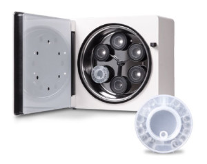
I followed up by asking about the specific components of the product itself and how it fits into a standard lab. Prof. Fey shared that their current system was composed of two parts. The ClinoStar, which consists of 6 clinostats wrapped up in a CO2 incubator and a tablet to run the software that regulates the speed of the clinostats, the temperature, and CO2 levels.
Also included are round disposable cell culture flasks, a bit like a petri dish that you can close, called ClinoReactors. The ClinoReactors contain their own humidification, so the ClinoStar can be run without humidity, which significantly reduces the risk of infections. The ClinoStar also has a built in UVC lamp to kill bacteria.
He did say that assays need to be changed, but CelVivo has published a collection of assays that have been tested and work well with their system. They system makes it is possible to accurately measure the amount of cells, DNA, and protein in the clusters simply by measuring their shadow area. That means that it is easy to standardize experiments without using any material.
I then asked if he could walk us through an organoid workflow using the ClinoStar. He said that if you are working with a cell line, the workflow is to grow and collect your cells from classical 2D cultures as usual. Then the cells can either be put directly into a culture vessel or you can form small clumps, then put them into the culture vessel and rotate them. Primary or stem cells can be treated the same way. The media needs to be changed every second day or so, but over the course of 2-3 weeks, little pieces of mimetic tissue form, which can then be used for research. If you are working from biopsies, then the cells need to be dispersed from the biopsy, or you can just produce very small pieces and proceed from there. These are usually functional sooner, but it depends on your system and how you start the clusters.
I asked where he thought that the ClinoStar has the most impact today. Prof. Fey said that the most exciting thing for him is to see the product used for many different applications. It is being used in drug discovery, testing medicinal plant extracts, epigenetics and cancer research, or for diabetes research, where the product originated from. Still others are working with bone growth or tissue aging. It has proven to be really useful in many different experimental settings and in each and every situation, it is providing data valuable for medical research.
I closed by asking if there was anything else that he would like to add for listeners. He said that he couldn’t go into any specific details, but he did say that they are planning to make the products even easier to use and more automatic. Also they are planning to be able to collect more data directly from the cultures.
Please visit ClinoStar, to learn more.