Launch of IncuCyte SX1 Model Expands Live-Cell Analysis Accessibility and Options
Cell models and cell systems have continued to evolve to meet research and drug discovery demands. Researchers are looking to increase physiological relevance of their cell models to ensure potential therapeutics have a higher chance of being both efficacious and safe. This desire has prompted many researchers to move from simpler recombinant cell systems to complex primary cell or stem-cell derived cell-based models. Co-cultures, multi-cultures and tissue organoid models offer significant promise, but also pose a greater challenge than the typical cell culture workflow. In addition, these cells and cell models tend to be more sensitive to minor deviations in their culture environment and as a result often have specialized requirements.
In addition to special culture conditions, advanced cell models also require enhanced instrumentation. One of the main advantages of using advanced cell models is their ability to provide detailed information on cellular changes in response to treatments or other external stimuli. This necessitates instrumentation that can maintain cell viability while being able to capture important data points throughout the culture period. As a result, imaging systems are needed that reliably analyze dynamic changes of cells in a stable, physiological environment throughout the entire experiment. Because these models provide insight into many complex interactions, the best opportunity for full understanding is to monitor them around the clock.
Live-Cell Analysis
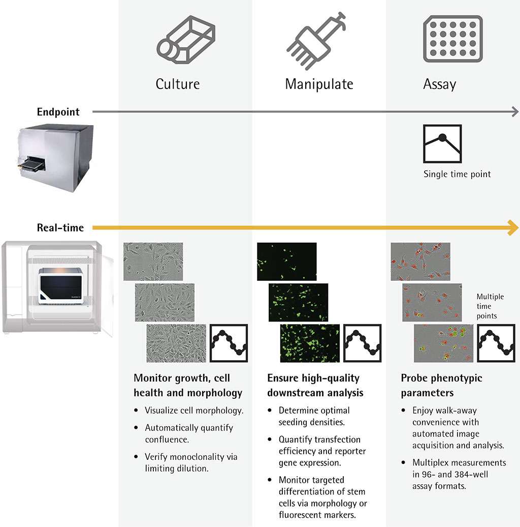
One method to manage the kind of information generated by advanced cell models is to use live-cell analysis. Live-cell analysis provides around the clock monitoring of cells and allows for repeated measurements of the same population of cells. Real-time cellular analysis allows cells to be evaluated to see how they grow, interact, and evolve over time, which cannot be achieved with single end-point evaluation. Cells can be analyzed for days, weeks or even months as they are maintained in the stable environment of a tissue culture incubator. This provides access to more information about the cell culture and models when compared with end-point workflows (Figure 1).

IncuCyte® Live-Cell Analysis Imaging Systems
Sartorius’ IncuCyte® S3 Live-Cell Analysis System has been very popular for live-cell imaging and analysis with over 2,500 peer-reviewed articles demonstrating unique applications of the IncuCyte with more being developed regularly. The IncuCyte S3 can accommodate multiple experiments in parallel with up to six microplate experiments at a time. This is an ideal system for a lab that has a substantial workload with many users performing live-cell analysis on a daily or weekly basis. However, it may not be a perfect fit for smaller labs or individual users.
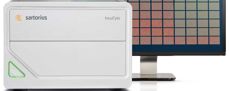
To expand available options for live-cell analysis, Sartorius launched their new entry level IncuCyte® SX1 Live-Cell Analysis System at ISSCR last month. This new model offers the same information-rich analysis and streamlined user experience as the IncuCyte S3, but has been designed to meet the specific workload and workflow needs of labs that might be in early phases of their live-cell analysis research program, or have fewer users. This expanded access permits live-cell, automated image acquisition and analysis at any scale.
Key Benefits of IncuCyte Live-Cell Analysis Systems
Supports the Entire Workflow
Culture Monitoring
All models of the IncuCyte Live-Cell Analysis System permit researchers to monitor the growth, morphology and health of their cells throughout the entire culture period. This includes the ability to visualize healthy cell morphology, automatically quantify confluence and verify monoclonality using limiting dilution. Cells are protected during this process as analysis is performed under uninterrupted incubation provided by a standard tissue culture incubator using IncuCyte’s non-perturbing reagent formulations.
Manipulating Cells in Culture
The system also ensures the quality of downstream analysis by determining optimal seeding densities, quantifying transfection efficiency and reporter gene expression, and monitoring targeted differentiation of stem cells via morphology or fluorescent markers.
Assaying Cells
The system permits kinetic assaying and the ability to probe phenotypic parameters, as well as analyze cell health, movement, morphology or function in response to treatments or manipulations.
Improving Productivity
Built in automation enables users to walk away while images are automatically acquired and analyzed. Measurements are available in 96- and 384- well assay formats.
Therapeutic Areas and Applications
As discussed, the IncuCyte has over 2,500 peer-reviewed articles discussing IncuCyte applications. With this prolific use, it is clear that researchers are using the IncuCyte to achieve better imaging and analysis of their advanced cell models and are also using the instrument to gain insights into new applications. This means that researchers are getting answers to unique scientific questions using kinetic image-based measurements as well as having the ability to design new experiments that were not previously possible.
This makes it possible to generate new insights quickly with a system designed for a range of cell types from proliferating tumor cells to non-adherent immune cells to sensitive primary cells. It also enables profiling of cell-specific and time dependent biological activity.
Ease of Use and Support
Sartorius develops applications from the ground up using appropriate reagents, cultureware, and purpose-built software to get a scientist from point A to point B with as little trial and error as possible. Once the best approach has been determined, they present the user with an end-to-end solution, including lab-tested protocols, and a guided software interface that streamlines the entire experience.
For more information please see IncuCyte portfolio of products
Related Content
Don’t miss our podcast interview and accompanying article, “How Live Cell Analysis Technology is Meeting the Needs of Ever Evolving Advanced Cell Models,” with Dr. Kimberly Wicklund, Head of Product Management for the IncuCyte® at Sartorius. We discussed how live cell analysis is meeting the needs of advanced cell models and how the launch of the new IncuCyte SX1 is providing scientists more options when it comes to using live cell analysis in their workflows.